
Artificial Intelligence (AI) is playing a transformative role in radiology, especially in CT (Computed Tomography) and MRI (Magnetic Resonance Imaging). As technology advances, AI is being used to improve the quality of medical images, streamline scan procedures, and reduce risks to patient safety — particularly those related to radiation exposure and contrast agents.
In this article, we explore how AI is reshaping modern medical imaging and what it means for patients undergoing MRI or CT scans in Paphos and across Cyprus.
Want to know more about MRI scans in Paphos? Everything you need to understand about the MRI process, safety measures, and preparation steps.
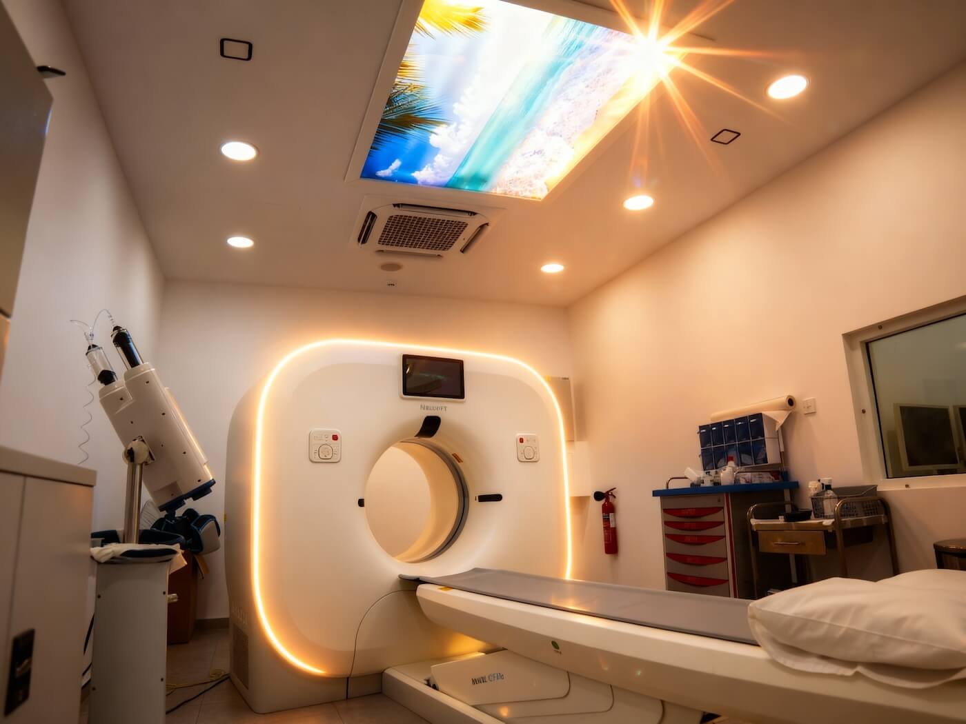
How AI Improves CT Imaging
CT scans are essential for diagnosing a wide range of conditions — from internal injuries and lung infections to abdominal and vascular issues. With the integration of AI, CT imaging is becoming faster, more accurate, and safer.
Key improvements include:
- Better patient positioning: AI algorithms can automatically align the patient and adjust table height, reducing radiation exposure and improving image accuracy.
- Optimised scan range: AI accurately defines the area to be scanned, avoiding unnecessary imaging and radiation.
- Personalised scan settings: Based on each patient’s size and condition, AI helps customise parameters to improve clarity and reduce human error.
- Contrast media optimisation: AI reduces the amount of contrast agent needed while maintaining image quality, minimising health risks.
- Improved image reconstruction: Deep learning models reduce noise and enhance clarity, allowing for high-quality images even with lower radiation doses.
These advancements mean patients can receive clearer results while being exposed to less radiation — a significant step forward in patient safety for CT scans in Paphos and beyond.
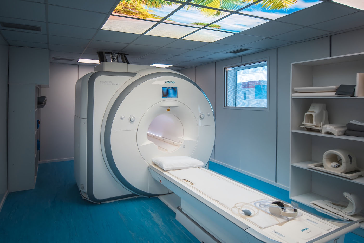
AI Innovations in MRI Imaging
MRI scans are known for their detailed images of soft tissues, making them crucial for diagnosing brain, spine, joint, and organ conditions. Traditionally, MRI can take longer to complete, leading to patient discomfort and motion-related image distortions.
AI is helping overcome these issues by:
- Speeding up scan times: AI-enhanced reconstruction allows high-resolution images to be captured in less time, reducing discomfort and motion artefacts.
- Reducing contrast agent use: AI can help reduce the need for gadolinium-based contrast agents (GBCAs) by up to 90%, which is beneficial for patients requiring repeated scans.
- Correcting motion distortions: Especially valuable in children and cardiac imaging, AI detects and adjusts for patient movement, improving accuracy.
These improvements make MRI scans in Paphos not only faster and more comfortable, but also safer for those who need regular imaging.
Current Challenges and Future Outlook
While AI offers promising benefits, there are still challenges to address:
- Generalisability: AI models often rely on specific scanners or protocols, which can limit their use across different facilities.
- Transparency: Many AI systems operate like “black boxes,” making it difficult for clinicians to understand how results are generated.
- Validation and regulation: Widespread clinical adoption will require more testing, validation across diverse patient groups, and clear regulatory guidelines.
To fully unlock AI’s potential, the focus must shift toward explainable AI, standardised datasets, and collaborative efforts between developers, radiologists, and regulators.
What This Maeans for Patients
For patients undergoing CT or MRI scans in Cyprus, AI is quietly working in the background to improve their experience — from faster appointment times to safer scan protocols. These technologies are supporting doctors with more reliable diagnostic tools, contributing to earlier detection and better treatment planning.
As AI continues to evolve, it is expected to become a core part of radiological workflows, improving outcomes while keeping patient safety at the forefront.
Published by Medscan Radiology Center | Source: European Radiology Experimental